
Human Organ Atlas: HiP-CT imaging of a COVID-19 injured human lung using the ESRF-EBS
Human Organ Atlas: HiP-CT imaging of a COVID-19 injured human lung using the ESRF-EBS
 Seeing inside a COVID-19 injured lung using a new imaging technique – Hierarchical Phase-Contrast Tomography or ‘HiP-CT’. Performed at the ESRF-EBS 4th generation synchrotron in Grenoble
HiP-CT is a new technique that can hierarchically image intact whole human organs. Beginning with a scan of the whole organ at the resolution of a human hair (25μm/voxel), followed by zooming in to any area at a resolution of 1/10th a human hair (6μm/voxel), and finally zooming in again to a resolution where we can see single cells (1.5μm/voxel).
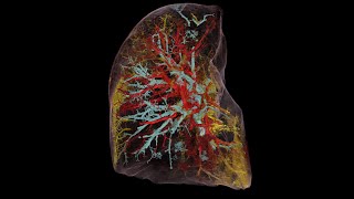
HiP-CT imaging of a whole left lung lobe from a 54yr old male donor showing injury due to COVID-19 using the new capabilities of ESRF-EBS for the Human Organ Atlas.
COVID-19 damages the lungs, causing changes in the structure of the lung itself. The lung is essential for oxygenating our blood and should be mostly filled with air. As we go through image slices of the lung you will see white, black and grey areas. The black areas are where air would be and the white and grey are the lung tissue and the blood vessels. In the COVID-19 case there is a lot more white and grey than in the healthy lung. As we zoom into this lung you can see how the air spaces are filled with a light grey colour indicating fluid and as we zoom in more you may notice how there seem to be many more dense and obvious blood vessels than in the healthy lung. Video produced by Paul Tafforeau.
Results for scientific and medical research and educational use only. HiP-CT and the Human Organ Atlas originated from a group trying to understand how COVID-19 injures our organs. The group are now developing HiP-CT to map our organs in health and disease to better understand them from a whole organ system down to the cellular level.
Please contact hipct.ucl@gmail.com for further information. Project Investigators/contributors: Peter D. Lee and Claire Walsh (UCL), Paul Tafforeau (ESRF), Danny Jonigk (Hannover), Maximilian Ackermann (Mainz), Will Wagner (Heidelberg), Joe Jacob and Simon Walker-Samuel (UCL), Mark Kuehnel and Christopher Werlein (Hannover), Alexandre Bellier (LADAF), and many others helping. We wish to thank ESRF for continuing to support the development of this programme (led by Paul Tafforeau, Beamtimes MD-1252 and MD-1290).
The authors wish to thank the various funders of the authors and this project, including: the Chan Zuckerberg Initiative DAF, an advised fund of Silicon Valley Community Foundation; the Royal Academy of Engineering; the MRC; the Wellcome Trust; the Deutsche Forschungsgemeinschaft; the National Institutes of Health (NIH); and the German Registry of COVID-19 Autopsies (supported by the German Federal Ministry of Health).
This is the best use of technology I’ve ever seen. Thank you Paul Tafforeau.
As a Radiology student, it’s about time to have something new, because the old black and white imagery makes it also hard to identify the disease or illness
Outstanding! Was the donor under mechanical ventilation?
It seems to me that a comparison of healthy vs covid vs covid + mechanical ventilation is in order.
Serdar hocanın müdavimleriyiz
where to download the 3D file?